Physical Description
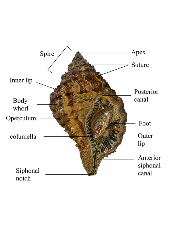
Figure a. Labelled diagram of C.
parthenopeum shell with the head-foot
retracted and the operculum closed.
Photo by Jacob Yeo, referenced from Wilson, 2002.
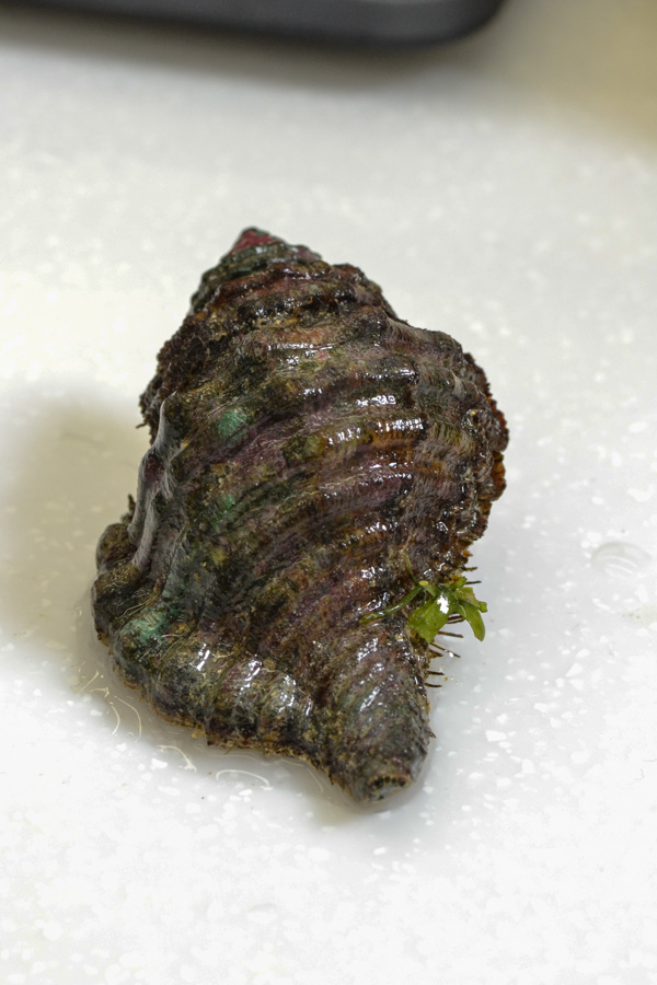
Figure b. Anterior view of the C.
parthenopeum shell.
Photo credit: Jacob Yeo
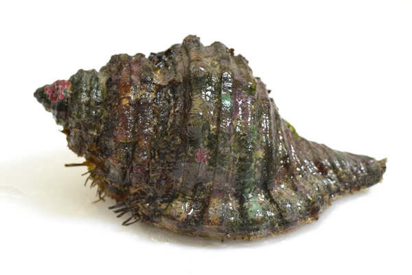
Figure c. Right side view of the C.
parthenopeum shell.
Photo credit: Jacob Yeo
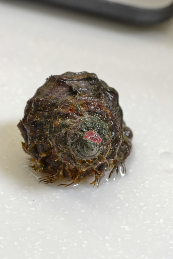
Figure d. Posterior view of the C.
parthenopeum shell.
Photo credit: Jacob Yeo
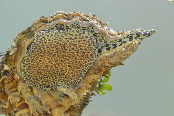
Figure e. Ventral view of C. parthenopeum,
showing distinct yellowish brown coloration
with dark brown spots on foot and inhalant
siphon.
Photo credit: Jacob Yeo
Max reported size: 150mm (ALA, 2014)
Cymatium (Monoplex) parthenopeum can be differentiated from others in the family by its broader shell. Its thick shell is covered by strong varices that are detailed by axial striae and nodulose spiral ribs. It also has a toothed lip with black and white markings around the aperture and a glazed, lirate columella. A long anterior canal forms the sprout. It has a light brownish yellow exterior covered with a thick and hairy periostracum (Wilson, 2002). The foot and inhalant siphon of the animal has a distinctive coloration of creamy greenish yellow with round dark brown spots. Compared to the adults, juveniles have a more swollen shell covered with long hairs forming dense rows (ALA, 2014).
|